

Skin Health Strategies to Reduce Inflammation With Functional Medicine
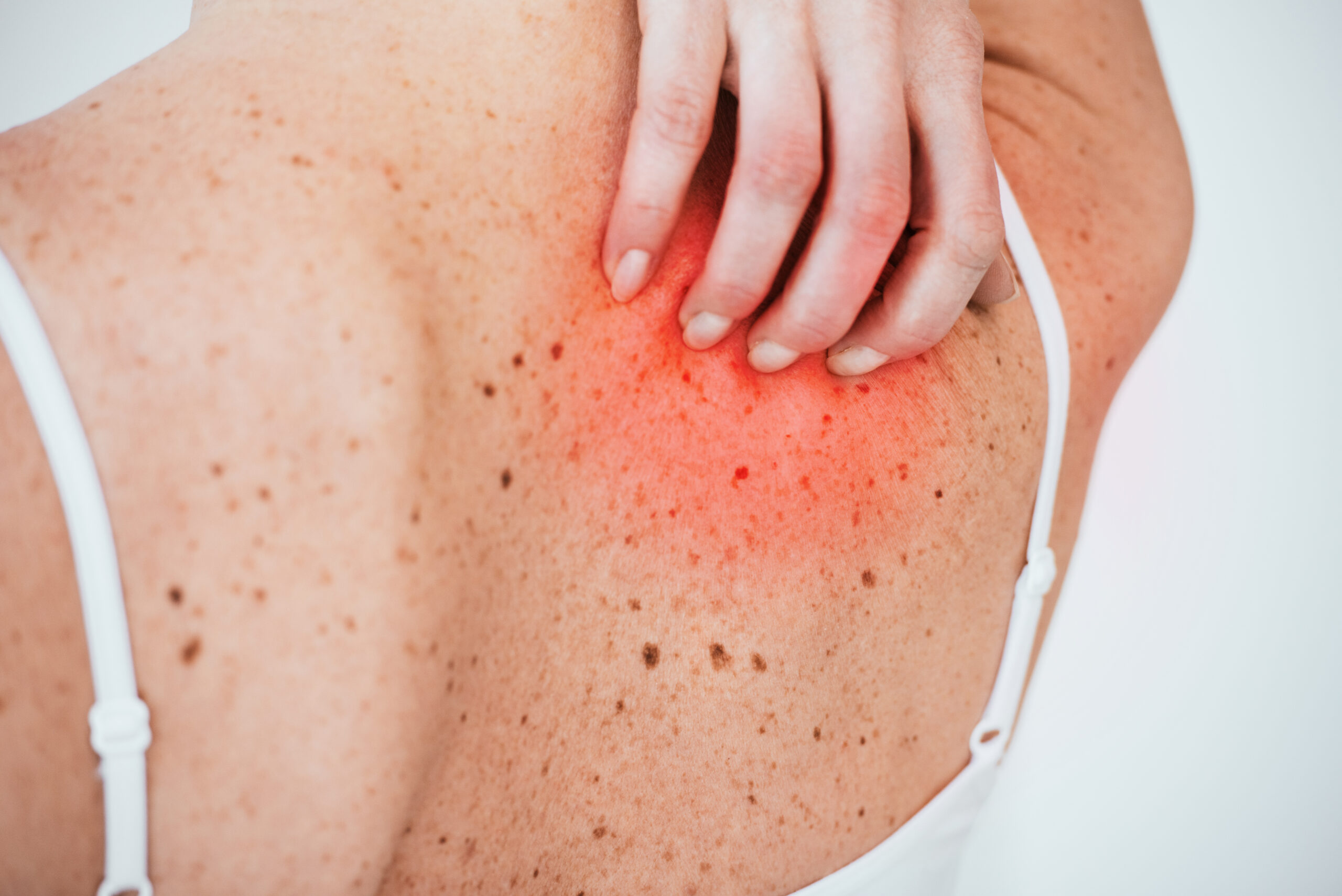
Uncover the benefits of functional medicine for improving skin health by reducing inflammation and achieving glowing skin.
Clinical Insight: Shave Skin Biopsy for Seborrheic Keratosis
Hello, I’m Dr. Alex Jimenez. It’s a privilege to share my clinical insights with you today. As a Doctor of Chiropractic (DC) and board-certified Family Nurse Practitioner (FNP-BC), my practice is deeply rooted in an integrative, functional medicine approach. Holding certifications as a Certified Functional Medicine Practitioner (CFMP), an Institute for Functional Medicine Certified Practitioner (IFMCP), and in Advanced T-Cell Neurology (ATN) and Cranio-cervical Instability and Trauma (CCST), I am dedicated to bridging the gap between conventional and complementary care.
At our practice, Injury Medical Clinic PA in El Paso, Texas, we have created a unique, multidisciplinary environment. I am honored to work alongside Dr. Maria Guadalupe Cardenas, MD, our Medical Director and Collaborative Physician. Dr. Cardenas is Board Certified in Internal Medicine and brings over 40 years of invaluable experience to our team. Her extensive knowledge and medical oversight (NPI #1164426749, Texas MD License #J2933) are foundational to our integrative model, allowing us to provide comprehensive, evidence-based care that addresses the whole person. Our collaborative setup ensures that patients receive the benefits of both advanced chiropractic and functional neurology alongside expert medical management.
This educational post will take you on a detailed journey through a common in-office procedure: the shave skin biopsy. We will explore not just the “how” but the critical “why” behind each step, grounding the procedure in established medical and physiological principles. I will discuss how we manage patient comfort, the specific techniques used to ensure an accurate diagnosis, and the importance of hemostasis. More importantly, I will connect this procedure to the broader philosophy of integrative care, explaining how something as specific as a skin biopsy fits within our holistic model of health, which includes chiropractic care, functional medicine, rehabilitation, and personalized wellness strategies.
Our goal is to demystify medical procedures and empower you with knowledge. By understanding the body’s intricate processes and the rationale behind our treatments, you become an active partner in your health journey.
Abstract: An Integrative Approach to Dermatological Procedures
This educational article provides a detailed, first-person narrative of a shave skin biopsy procedure for an inflamed seborrheic keratosis. Authored by Dr. Alex Jimenez, it explains the clinical reasoning and physiological principles behind each step, from patient assessment and local anesthesia to the biopsy technique and post-procedural care. The post emphasizes patient comfort, the role of local anesthetics like lidocaine with epinephrine, and the technique of creating an intradermal wheal to facilitate the procedure. It also situates the procedure within integrative healthcare, showing how a focused dermatological intervention fits into a broader care model. This model combines the expertise of Dr. Alex Jimenez (Chiropractic, Functional Medicine) and Dr. Maria Cardenas (Internal Medicine) to address not just the presenting symptom but the patient’s overall health, including musculoskeletal alignment, systemic inflammation, and metabolic function. The article highlights how chiropractic adjustments and functional medicine interventions can support the body’s healing processes, reduce systemic stress, and contribute to long-term wellness, even when managing a specific issue like a skin lesion.
Initial Patient Presentation and Clinical Assessment
Today, September 1, 2026, I consulted with a middle-aged gentleman who presented with a concern that had troubled him for the past month. He pointed to a skin lesion on the lateral aspect of his right hip, which he reported had become increasingly noticeable over the last four weeks. Beyond its appearance, the lesion had begun to cause discomfort, with signs of inflammation and intermittent pain.
During the physical examination, my primary goal was to characterize the lesion based on its morphology, location, and the patient’s reported history. The lesion presented with features highly suggestive of an inflamed seborrheic keratosis.
Understanding Seborrheic Keratosis
A seborrheic keratosis (SK) is one of the most common benign (non-cancerous) skin growths that affect adults. They often appear in middle age or later and can occur almost anywhere on the body, except the palms and soles. Clinically, they have a characteristic “stuck-on” appearance, as if a piece of wax or a crumb of brown sugar was pasted onto the skin. They can range from light tan to dark brown or black and often have a waxy, scaly, or slightly raised surface.
While SKs are benign, they can become problematic for several reasons:
- Irritation and Inflammation: Because they are raised, they can be easily irritated by clothing, friction, or accidental scratching. This mechanical irritation can trigger a localized inflammatory response, causing redness, swelling, tenderness, and even bleeding, as in our patient.
- Diagnostic Uncertainty: Although they have a classic appearance, an inflamed or irritated SK can sometimes mimic more serious skin conditions, most notably nodular melanoma, a type of skin cancer. The inflammation can obscure the typical features of the SK, creating what we call an “atypical presentation.”
- Patient Discomfort and Concern: The sudden change in a long-standing skin lesion—such as enlargement, inflammation, or pain—is a significant source of anxiety for patients, and rightly so. Any “changing mole” or lesion warrants a thorough evaluation.
Given the patient’s symptoms of recent enlargement, inflammation, and pain, a definitive diagnosis was essential. While my clinical impression strongly suggested an inflamed seborrheic keratosis, the only way to be 100% certain and rule out underlying malignancy is through histopathological examination. This requires obtaining a tissue sample for a pathologist to analyze under a microscope. Therefore, we decided to perform a shave skin biopsy. This procedure would serve a dual purpose: provide a definitive diagnosis while removing the bothersome lesion.
This initial assessment is a critical step in our integrative model. It’s not just about identifying a skin lesion; it’s about listening to the patient’s experience, understanding their concerns, and formulating a plan that addresses both the clinical need and the patient’s peace of mind. Under the collaborative guidance of Dr. Cardenas, our medical director, proceeding with a biopsy is the standard of care and aligns perfectly with our commitment to evidence-based practice.
Preparing for the Procedure: Anesthesia and Patient Comfort
The success and patient experience of any minor procedure are heavily dependent on meticulous preparation, particularly in managing and preventing pain. My philosophy, shared by our entire team at Injury Medical Clinic, is that medical care should be as comfortable and stress-free as possible. A patient who feels safe and experiences minimal discomfort is more relaxed, which physiologically contributes to a better outcome. The body’s stress response (the “fight-or-flight” mechanism) can increase blood pressure and heighten pain sensitivity, so mitigating this is a clinical priority.
Step 1: Antiseptic Preparation
Before any instrument touches the skin, ensuring a sterile field is paramount to prevent iatrogenic infection (an infection caused by medical examination or treatment).
- Action: I thoroughly cleansed the area over the lateral right hip, encompassing the lesion and a wide margin around it, using an alcohol prep pad.
- The “Why”: The skin is naturally colonized with a vast microbiome of bacteria, including species like Staphylococcus aureus and Streptococcus pyogenes. While these are often harmless on the skin’s surface, introducing them into the deeper dermal or subcutaneous layers via a needle or scalpel can lead to cellulitis or a localized abscess. Alcohol is a potent bactericidal agent that denatures these microorganisms’ proteins, reducing the bacterial load on the skin surface and minimizing infection risk.
Step 2: Topical Vapocoolant for Initial Anesthesia
The first moment of pain in this procedure is the needle stick itself. While it’s a minor sensation, it can trigger anxiety. To blunt this initial sting, I employed a topical anesthetic spray.
- Action: I administered a brief spray of Pain Ease® Mist, a vapocoolant, directly onto the target injection site.
- The “Why”: Vapocoolants like Pain Ease® contain volatile chemicals (such as ethyl chloride or a blend of tetrafluoroethane, pentafluoropropane, and tetrafluoropropene) that evaporate almost instantaneously upon contact with the skin. This rapid evaporation causes a significant and immediate drop in skin temperature. This intense cooling effect works via the “Gate Control Theory of Pain” (Melzack & Wall, 1965). The theory posits that non-painful sensory information (in this case, the sensation of cold) can “close the gates” to painful stimuli. Cold signals travel along large, fast-conducting A-beta nerve fibers, which inhibit the transmission of pain signals carried by smaller, slower A-delta and C fibers at the spinal cord’s dorsal horn. Essentially, the brain is “distracted” by the intense cold, so the sharp pain of the needle prick is perceived as duller or not at all. This simple, non-invasive step significantly improves the patient’s comfort from the very outset.
Step 3: Injectable Local Anesthesia – The Core of Pain Control
With the initial needle sensation minimized, the next and most crucial step is to deliver a local anesthetic to numb the entire procedural area.
- Action: I used a very fine 30-gauge, half-inch needle to insert into the subcutaneous tissue adjacent to the lesion quickly. I then injected approximately 1 milliliter (mL) of 1% lidocaine with epinephrine.
- The “Why”: This choice of anesthetic and needle is deliberate and based on several physiological principles:
-
- Needle Gauge: A 30-gauge needle is one of the smallest-diameter needles commonly used in clinical practice. A smaller diameter means less tissue displacement and fewer pain receptors stimulated upon entry, further minimizing discomfort.
- Lidocaine: Lidocaine is the “workhorse” of local anesthetics. It is an amide-type anesthetic that blocks voltage-gated sodium channels in the neuronal cell membrane. Nerve impulses (including pain signals) are transmitted as electrical signals called action potentials, which depend on the rapid influx of sodium ions into the neuron. By blocking these sodium channels, lidocaine prevents the nerve from depolarizing and firing, thus stopping the transmission of pain signals from the procedural site to the brain. The area becomes profoundly numb within minutes.
- Epinephrine: The addition of epinephrine, a potent vasoconstrictor, is critically important for two reasons. First, it constricts the small blood vessels (arterioles and venules) at the injection site. This reduces local blood flow, which slows the rate at which lidocaine is absorbed into the systemic circulation. The result is that the lidocaine remains concentrated at the site of injection for a longer period, significantly prolonging the duration of anesthesia. This keeps the patient comfortable throughout the procedure and afterward. Second, the vasoconstriction provides excellent hemostasis (control of bleeding). By clamping down on the local blood supply, it minimizes bleeding during the biopsy itself, which is essential for maintaining a clear view of the lesion and ensuring a clean, precise excision.
After the initial subcutaneous depot injection, I asked the patient, “Does that hurt?” He responded, “No.” This immediate feedback is crucial. It confirms that our pain management strategy is effective and builds the patient’s trust and confidence.
This multi-pronged approach to anesthesia—combining psychological reassurance, topical cooling, and a carefully chosen injectable agent—is a hallmark of patient-centered care. It turns a potentially stressful event into a manageable, comfortable experience.
The Biopsy Technique: Creating the Intradermal Wheal and Performing the Shave
With the area profoundly anesthetized, the next phase involves preparing the lesion for removal and performing the shave biopsy. The technique I used is designed to optimize the specimen for the pathologist while ensuring clean, efficient removal.
Redirecting the Needle to Create an Intradermal Wheal
Instead of removing the needle after the initial subcutaneous injection, I carefully redirected it. This technique minimizes skin punctures and associated discomfort.
- Action: Without pulling the needle out of the skin, I angled it horizontally and advanced it into the intradermal space directly beneath the seborrheic keratosis. I then slowly injected more of the lidocaine with epinephrine solution.
- The “Why”: This step is perhaps the most elegant and technically important part of the preparation. Injecting fluid into the dermis—the layer of skin just below the epidermis—creates a firm, elevated, pale blister known as a wheal. You can see this happen in real time as the skin blanches and rises. This intradermal wheal serves several critical functions:
-
- Elevates and Presents the Lesion: The fluid pressure from the wheal physically lifts the lesion, pushing it up and away from the underlying subcutaneous tissue. This “presents” the lesion beautifully, making it a much easier target to excise. It transforms a relatively flat or slightly raised area into a distinct, taut dome.
- Creates a Plane of Dissection: The fluid-filled wheal creates a clear, safe plane for the biopsy blade. It buffers the base of the lesion from deeper tissues, reducing the risk of cutting too deep and causing unnecessary tissue damage or scarring.
- Enhances Anesthesia: By injecting the anesthetic directly into the dermis where the nerve endings are most concentrated, it ensures profound local numbness precisely where the cut will be made. I checked in with the patient again, asking if this part was stinging (as intradermal injections sometimes can). His “No” was reassuring.
- Improves Hemostasis: The epinephrine in the solution, now distributed throughout the dermal capillaries beneath the lesion, provides maximum vasoconstriction exactly where it is needed most. This creates a relatively bloodless field for the biopsy.
As I injected the solution, I noted, “You’ll see that you’re developing an intradermal wheal. And it’s actually elevating the lesion toward us, so that presents it to us, so that we can very easily and quickly perform a shave biopsy.”
Performing the Shave Biopsy
With the lesion perfectly presented and the area numb, it was time for the excision. A shave biopsy involves using a sharp blade to remove the lesion at a level parallel to the skin surface.
- Action: Using a sterile DermaBlade, I began the incision approximately two millimeters lateral to the visible edge of the lesion. With a smooth, sawing motion, I advanced the blade horizontally through the dermis, staying within the plane created by the intradermal wheal. I continued this motion through to the other side of the lesion, ensuring the entire growth was removed in one continuous piece.
- The “Why”:
-
- Choice of Instrument: A DermaBlade or a similar tool (like a #15 scalpel blade held flat) is ideal for shave biopsies. It is flexible and extremely sharp, allowing precise control over cut depth.
- Starting Lateral to the Lesion: Beginning the cut just outside the lesion’s border ensures that a small margin of normal-appearing skin is included with the specimen. This is crucial for the pathologist, as it allows them to see the transition from normal tissue to the lesion, which can be important for diagnosis, especially if there is any suspicion of malignancy.
- Horizontal, Sawing Motion: A gentle, back-and-forth sawing motion allows the blade to glide through the tissue with minimal downward pressure. This prevents the operator from inadvertently cutting too deep. The goal is a superficial shave that removes the epidermis and a portion of the upper dermis.
- One Continuous Piece: Removing the lesion in a single piece is vital for accurate histopathological interpretation. If the specimen is fragmented, the pathologist may have difficulty assessing its architecture, margins, and depth.
After completing the pass, I examined the site. The lesion was completely removed, leaving a shallow, clean wound base. I noted, “All the way through, no lesion is left there on the skin.” I immediately placed the specimen in a properly labeled formalin jar and prepared it for transport to the pathology lab.
This precise, methodical approach ensures we obtain the best possible diagnostic sample while minimizing tissue trauma and optimizing the patient’s cosmetic outcome.
Post-Procedure Care: Hemostasis and Wound Management
Once the biopsy specimen has been secured, the final and equally important step is to manage the biopsy site. The primary goals are to achieve immediate hemostasis (stopping the bleeding) and to create an environment conducive to proper wound healing.
Achieving Hemostasis with Aluminum Chloride
Even with epinephrine’s vasoconstrictive effects, a shave biopsy creates a raw dermal surface with open capillaries that ooze blood. Active hemostasis is required.
- Action: I took a sterile gauze pad and saturated it with aluminum chloride solution. I then applied the saturated gauze directly to the wound base with firm, continuous pressure.
- The “Why”: Aluminum chloride is a powerful styptic agent, meaning it stops bleeding. It works differently than epinephrine. Aluminum chloride is a chemical cauterant that acts as an astringent. When it contacts blood and tissue proteins, it causes them to precipitate and denature. This creates a physical plug of coagulated protein that seals the ends of the severed capillaries, effectively stopping the bleeding.
-
- Mechanism of Action: The trivalent aluminum ions (Al³+) interact with proteins like albumin in the blood and structural proteins in the tissue, causing them to clump together and form a mechanical barrier. This is a rapid, effective way to control the superficial, oozing-type bleeding characteristic of a shave biopsy wound.
- Comparison to Other Agents: While other hemostatic agents exist (e.g., silver nitrate, which can cause skin staining, or electrocautery, which involves heat), aluminum chloride is often preferred for superficial shave biopsies because it is effective, easy to apply, and does not typically cause discoloration.
After a few moments of pressure, I removed the gauze. As expected, the bleeding had completely stopped. The wound base was clean and dry. I noted, “The bleeding has stopped.”
Wound Dressing and Patient Instructions
With hemostasis achieved, the final step is to protect the wound.
- Action: The site was covered with a sterile dressing—typically an antibiotic ointment (like bacitracin or a simple petrolatum-based ointment) and a sterile adhesive bandage.
- The “Why”:
-
- Antibiotic/Petrolatum Ointment: Applying an ointment serves several purposes. It keeps the wound moist, which has been shown to accelerate healing and reduce scarring compared to letting a wound dry out and form a hard scab (Winter, 1962). It also provides a barrier against environmental bacteria, further reducing infection risk. While topical antibiotics were once standard, many guidelines now favor plain petrolatum to avoid the risk of contact dermatitis or antibiotic resistance. The primary goal is to maintain a moist, occlusive environment.
- Sterile Bandage: The bandage protects against friction and contamination, absorbs any minor residual seepage, and keeps the ointment in place.
The patient was then given clear, simple instructions for wound care at home:
- Keep the bandage on and dry for the first 24 hours.
- After 24 hours, remove the bandage. Gently cleanse the area daily with mild soap and water.
- After cleansing, apply a thin layer of petrolatum ointment, then apply a new bandage.
- Continue this routine until the wound has fully epithelialized (i.e., new skin has covered the surface), which typically takes 7 to 14 days for a shave biopsy of this nature.
- We also instructed the patient to watch for signs of infection, such as increasing redness, swelling, pain, warmth, or purulent drainage, and to contact our clinic immediately if any of these occur.
Finally, we informed the patient that we would contact them with the pathology results as soon as they were available, usually within 5-10 business days. This communication is key to closing the loop and alleviating any lingering anxiety about the lesion.
This comprehensive approach to post-procedural care helps the patient heal quickly and without complications, reinforcing a positive experience at our clinic.
The Healing Diet: Combat Inflammation, Embrace Wellness- Video
The Integrative Framework: Connecting the Biopsy to Whole-Person Health
Performing a shave biopsy is a discrete, technical procedure. However, within the walls of Injury Medical Clinic PA, it is never just an isolated event. It is one data point in a much larger, integrated picture of the patient’s health. This is where the synergy between my role as a Doctor of Chiropractic and Functional Medicine Practitioner and Dr. Cardenas’s expertise in Internal Medicine truly shines. Our model is built on the understanding that the body is an interconnected system, and a skin lesion can reflect deeper systemic processes.
Systemic Inflammation and Skin Health
The patient’s lesion was described as “inflamed.” While local irritation from clothing is a likely culprit, we also consider an exaggerated inflammatory response due to an underlying pro-inflammatory state. In functional medicine, we investigate the root causes of chronic inflammation, which can manifest in diverse ways, including on the skin.
- Potential Contributors to Systemic Inflammation:
-
- Diet: A diet high in processed foods, sugar, and inflammatory fats (like omega-6 fatty acids) and low in anti-inflammatory nutrients (like omega-3s and phytonutrients) can fuel chronic, low-grade inflammation throughout the body (Sears & Ricordi, 2011).
- Gut Health: The gut is a major regulator of the immune system. A condition known as intestinal permeability, or “leaky gut,” allows undigested food particles and bacterial endotoxins (like lipopolysaccharide, LPS) to enter the bloodstream, triggering a body-wide inflammatory cascade that can affect the skin.
- Stress: Chronic psychological stress leads to sustained high levels of the hormone cortisol. While cortisol has anti-inflammatory effects in the short term, chronically elevated levels can lead to cortisol resistance and immune dysregulation, ultimately promoting inflammation (Morey et al., 2015).
- Metabolic Dysfunction: Conditions like insulin resistance and metabolic syndrome are fundamentally inflammatory states.
As part of our integrative follow-up, we would discuss these factors with the patient. A functional medicine workup might include advanced testing to assess inflammatory markers (like hs-CRP), nutrient status, and gut health. Based on these findings, we could recommend personalized dietary changes, targeted supplementation (e.g., curcumin, fish oil), and stress management techniques. This approach doesn’t just treat the skin lesion; it aims to lower the patient’s overall inflammatory burden, potentially reducing the risk of future inflammatory issues, both dermatological and systemic.
The Role of Chiropractic Care in Systemic Well-Being
At first glance, chiropractic care might seem unrelated to a skin biopsy. However, from an integrative and neurological perspective, it is profoundly connected to the body’s ability to heal and regulate itself.
- Neuro-Immune Modulation: The nervous, endocrine, and immune systems are intricately linked in a complex communication network. Chiropractic adjustments are designed to correct vertebral subluxations—misalignments or dysfunctional segments of the spine that can interfere with nerve function. Research suggests that by restoring proper motion and signaling within the nervous system, chiropractic care can modulate the autonomic nervous system (ANS) (Kovanur-Sampath et al., 2017).
- Balancing the Autonomic Nervous System: The ANS has two main branches: the sympathetic (“fight-or-flight”) and the parasympathetic (“rest-and-digest”). Chronic stress, pain, and inflammation can lead to sympathetic dominance. This state is associated with poor tissue healing, increased inflammation, and heightened pain perception. Chiropractic adjustments, particularly of the upper cervical and sacral regions, have been shown to promote a shift toward parasympathetic activity. This “rest-and-digest” state supports healing, tissue repair, and immune regulation. By helping the patient move out of chronic stress, we create a physiological environment more conducive to healing the biopsy site and managing overall inflammation.
- Pain Management and Structural Integrity: Our patient presented with the lesion on his hip. In our comprehensive assessment, we would also evaluate his biomechanics. Is there a pelvic imbalance or a gait abnormality that is causing his clothing to rub preferentially on that spot? Correcting underlying musculoskeletal imbalances through chiropractic adjustments and rehabilitative exercises could address the mechanical source of the irritation that caused the lesion to become inflamed in the first place. This is a classic example of looking for the “why behind the why.” The inflammation was caused by friction, but what caused the friction? Addressing this root structural issue is key to preventing recurrence.
A Collaborative, Multidisciplinary Approach
This is the essence of our practice. The shave biopsy, overseen by Dr. Cardenas and performed with my training as a nurse practitioner, addresses the immediate medical need according to the highest standards of care. We send the tissue for pathological diagnosis, a cornerstone of allopathic medicine.
Simultaneously, we use this event as an entry point to a broader conversation about health:
- Medical Oversight (Dr. Cardenas): As Medical Director, Dr. Cardenas ensures all procedures and treatment plans are medically sound, safe, and appropriate. Her four decades of experience in internal medicine provide an invaluable depth of knowledge for diagnosing and managing complex cases and comorbidities.
- Procedure and Functional Medicine (Dr. Jimenez, as FNP & CFMP): I perform the procedure and also lead the investigation into the underlying functional imbalances. Are there dietary triggers? Is there a gut-skin axis issue? Is chronic stress a factor?
- Chiropractic and Neurological Care (Dr. Jimenez, as DC): I assess and address the structural and neurological components. Is there a subluxation affecting autonomic tone? Is a biomechanical fault contributing to the initial irritation?
This integrated team approach lets us see the patient not as a skin lesion on a hip, but as a whole person whose skin communicates something about their overall health. By combining the diagnostic precision of conventional medicine with the root-cause focus of functional medicine and the neuro-structural support of chiropractic care, we can provide a truly holistic and powerfully effective form of healthcare. We don’t just remove the lesion; we partner with the patient to build a foundation for more resilient and vibrant health in the long term.
References
- Kovanur-Sampath, K., Mani, R., Cotter, J., & Tumilty, S. (2017). Changes in biochemical markers following spinal manipulation: a systematic review and meta-analysis. Musculoskeletal Science and Practice, 29, 120–131. https://doi.org/10.1016/j.msksp.2017.04.004
- Melzack, R., & Wall, P. D. (1965). Pain mechanisms: A new theory. Science, 150(3699), 971–979. https://doi.org/10.1126/science.150.3699.971
- Morey, J. N., Boggero, I. A., Scott, A. B., & Segerstrom, S. C. (2015). Current directions in stress and human immune function. Current Opinion in Psychology, 5, 13–17. https://doi.org/10.1016/j.copsyc.2015.03.007
- Sears, B., & Ricordi, C. (2011). Anti-inflammatory nutrition as a pharmacological approach to treating diabetes. Industrial & Engineering Chemistry Research, 50(15), 8993-9000. This reference may be better represented by a primary research article on diet and inflammation, such as: Christ, A., Lauterbach, M., & Latz, E. (2019). Western diet and the immune system: an inflammatory connection. Immunity, 51(5), 794-811. https://doi.org/10.1016/j.immuni.2019.09.020
- Winter, G. D. (1962). Formation of the scab and the rate of epithelization of superficial wounds in the skin of the young domestic pig. Nature, 193(4812), 293–294. https://doi.org/10.1038/193293a0
SEO Tags: shave biopsy, seborrheic keratosis, integrative medicine, functional medicine, chiropractic care, local anesthesia, lidocaine with epinephrine, intradermal wheal, hemostasis, aluminum chloride, skin health, systemic inflammation, Dr. Alex Jimenez, Dr. Maria Cardenas, El Paso TX, multidisciplinary care, pain management, vapocoolant, wound healing, patient-centered care, autonomic nervous system, vertebral subluxation, personal injury doctor El Paso, injury medical clinic
Comments are closed.