
Restorative Injection Therapy in Treating Musculoskeletal Pain
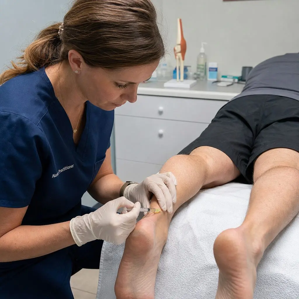
Find out how restorative injection therapy for musculoskeletal pain solutions can help you regain mobility and comfort.
Abstract
Hello, I’m Dr. Alex Jimenez. Welcome to this educational exploration of cutting-edge regenerative medicine and integrative care. In this detailed post, we will embark on an in-depth journey into the complex world of chronic musculoskeletal conditions, with a particular focus on tendinopathies like Achilles tendonitis. We will dissect the physiological processes that lead to these challenging injuries, contrasting the outdated concept of “tendonitis” with the more accurate model of “tendinosis,” which describes a degenerative, non-inflammatory state. Drawing from the latest findings of leading researchers, we will meticulously examine advanced, evidence-based treatment modalities, including Prolotherapy, Platelet-Rich Plasma (PRP), Extracorporeal Shockwave Therapy (ESWT), and amniotic tissue allografts. We will delve into the scientific rationale behind each protocol—from the precise concentration of dextrose in Prolotherapy to the cellular mechanisms activated by PRP and the mechanical forces of ESWT.
Furthermore, this post will illuminate the pivotal role of an integrative care model. I will explain how our practice, Injury Medical Clinic, synergizes the expertise of chiropractic care, functional medicine, and rehabilitation under my direction with the crucial medical oversight provided by our Medical Director and Collaborative Physician, Dr. Maria Guadalupe Cardenas, MD. This multidisciplinary framework ensures a comprehensive, patient-centered approach, addressing not just the localized injury but the systemic factors influencing recovery. We will explore how we combine these advanced injections and therapies with foundational chiropractic adjustments, nutritional support, and targeted rehabilitation to create a robust, holistic pathway to healing for our patients in El Paso, Texas, and beyond.
Our Integrative Approach: A Synergy of Expertise
Before we delve into the specifics of regenerative therapies, I believe it’s essential to set the stage by explaining the unique clinical model we’ve cultivated here at Injury Medical Clinic. Our philosophy is rooted in the understanding that the human body is an interconnected system, and treating it effectively requires a collaborative, multifaceted approach.
I am Dr. Alex Jimenez, and my journey in healthcare has led me to embrace a wide spectrum of disciplines, earning credentials as a Doctor of Chiropractic (DC), an Advanced Practice Registered Nurse (APRN) with a Family Nurse Practitioner Board Certification (FNP-BC), and certifications in Functional Medicine (CFMP, IFMCP), among others. This diverse background allows me to view patient health through multiple lenses—structural, neurological, biomechanical, and biochemical.
However, the cornerstone of our comprehensive care model is our collaboration with Dr. Maria Guadalupe Cardenas, MD. Dr. Cardenas is a highly respected physician, board-certified in Internal Medicine, with an impressive career spanning over 40 years. She serves as our Medical Director and Collaborative Physician, bringing a wealth of medical knowledge and diagnostic acumen to our practice. Her NPI number is 1164426749, and she is licensed to practice in Texas under license #J2933.
This partnership between a Doctor of Chiropractic and a Medical Doctor is fundamental to our practice’s identity. It allows us to operate as a true multidisciplinary clinic. Here’s how this synergy benefits our patients:
- Comprehensive Diagnostics: A patient presenting with chronic heel pain, for instance, receives a dual assessment. I conduct a thorough biomechanical and musculoskeletal evaluation, assessing gait, spinal alignment, and joint function. Simultaneously, under Dr. Cardenas’s medical direction, we can rule out or identify underlying systemic conditions like metabolic disorders, autoimmune diseases, or vascular issues that could be contributing to the symptoms.
- Safe and Medically Supervised Procedures: When we discuss advanced treatments like Prolotherapy or PRP injections, these are medical procedures. Dr. Cardenas provides the essential medical oversight required to perform these therapies safely and effectively. Her involvement ensures that we adhere to the highest standards of medical practice, from patient selection and contraindication screening to the sterile preparation and administration of injectables.
- Integrated Treatment Planning: Our patient care plans are not developed in a silo. Dr. Cardenas and I, along with our team of rehabilitation specialists and nutritionists, collaborate to create a unified strategy. A plan for Achilles tendinosis might include:
-
- Medical Intervention: An initial diagnostic ultrasound and, if necessary, a regenerative injection like Prolotherapy, overseen by Dr. Cardenas.
- Chiropractic Care: I would provide specific adjustments to the foot, ankle, and even the lumbar spine and pelvis to correct biomechanical imbalances and improve nerve function to the lower extremity.
- Functional Medicine: We would investigate and address potential nutritional deficiencies or inflammatory triggers that could be hindering tissue repair.
- Rehabilitation: Our physical therapy team would guide the patient through a carefully phased program of eccentric exercises and functional movements.
This integrated model ensures that we are not just chasing symptoms. We are addressing the root causes of dysfunction from every possible angle, providing a level of care that is not possible within a single-discipline practice. It is this collaborative spirit that defines our work at Injury Medical Clinic and Mission Plaza Injury Medical Clinic, allowing us to confidently tackle complex cases, including personal injury and chronic pain syndromes.
Rethinking Tendon Injuries: From “Tendonitis” to “Tendinosis”
For decades, the medical community used the term “tendonitis” to describe pain in a tendon. The “-itis” suffix implies inflammation. When a patient came in with a painful Achilles, elbow, or shoulder, the default diagnosis was inflammation of the tendon. Consequently, the standard treatment revolved around anti-inflammatory approaches: rest, ice, and non-steroidal anti-inflammatory drugs (NSAIDs) like ibuprofen or naproxen. If that failed, a corticosteroid injection was often the next step.
However, over the past 20-30 years, a paradigm shift has occurred, driven by meticulous research involving histology (the microscopic study of tissues). Groundbreaking work by researchers like Khan et al. (2002) has shown that in cases of chronic tendon pain, what we are actually seeing is not an inflammatory process but a degenerative one. When biopsies of these chronically painful tendons are examined under a microscope, they don’t show the classic inflammatory cells (like neutrophils and macrophages) that you would expect in an “-itis” condition.
Instead, what they reveal is a state of disrepair and degeneration, a condition more accurately termed “tendinosis.”
The Histopathology of Tendinosis
Let’s visualize what a healthy tendon looks like at the microscopic level versus a tendon in a state of tendinosis.
- Healthy Tendon: Imagine a thick, strong rope made of countless smaller fibers, all perfectly aligned, parallel, and tightly packed. This is a healthy tendon. It’s composed primarily of Type I collagen, which is incredibly strong and resilient. The collagen fibers are organized into neat bundles (fascicles), and the cells (tenocytes) are elongated and sit in orderly rows between these fibers. There is a well-organized blood supply and nerve network. This structure is designed to withstand immense tensile forces.
- Tendinopathic Tendon (Tendinosis): Now, imagine that same rope has become frayed, disorganized, and weak. This is tendinosis. The key features we observe are:
-
- Collagen Disarray: The beautiful, parallel alignment of Type I collagen fibers is lost. The fibers become thin, fragmented, and haphazardly arranged.
- Shift in Collagen Type: There’s a significant increase in the weaker, more fragile Type III collagen. This is the type of collagen the body lays down quickly during the initial phases of wound healing, almost like a temporary patch. In a healthy healing process, this Type III collagen is remodeled into strong Type I collagen. In tendinosis, this remodeling process fails, leaving the tendon structurally compromised.
- Angiofibroblastic Hyperplasia: This is a hallmark of tendinosis. It sounds complex, but it simply means there’s an abnormal proliferation of both blood vessels (angio) and fibroblasts (fibroblastic). However, these new blood vessels are not healthy and functional; they are leaky and disorganized. They are often accompanied by a disorganized ingrowth of nerve fibers, which is believed to contribute to the chronic pain associated with the condition (Alfredson, 2005).
- Increased Ground Substance: The “glue” that holds the collagen fibers together, known as the extracellular matrix or ground substance, increases. It becomes more gelatinous and less organized, further weakening the tendon’s structure.
This understanding is a game-changer. If the problem isn’t inflammation but degeneration, then treatments focused solely on reducing inflammation (like NSAIDs and corticosteroids) are not only missing the point but can be actively harmful. Corticosteroids, for example, are known to be catabolic, meaning they can break down tissue. Injecting a corticosteroid into a tendon that is already degenerating can further weaken it, increasing the risk of a complete rupture (Coombes et al., 2010).
This is why we’ve moved towards therapies that aim to regenerate the tissue and restart the healing process. Our goal is to shift the tendon from a degenerative state of tendinosis back into a proper, organized healing cascade.
The Critical Role of Immobilization and Offloading in Tendon Healing
Before we even consider advanced injections, we must address the fundamental biomechanical environment of the injured tendon. A tendon cannot heal if it is subjected to constant, repetitive strain that exceeds its current capacity. This is a non-negotiable principle of tissue healing.
Let’s use our primary example: Achilles tendinosis. The Achilles tendon is the largest and strongest tendon in the body, connecting the calf muscles (gastrocnemius and soleus) to the heel bone (calcaneus). Every time you walk, run, or jump, this tendon is loaded. In a healthy state, it acts like a powerful spring, storing and releasing energy. But in tendinosis, this normal loading becomes injurious, perpetuating the degenerative cycle.
Why a Walking Boot is Essential
This brings us to a crucial, and often resisted, part of the initial treatment protocol: immobilization with a walking boot.
From my clinical experience, patients often balk at the idea of wearing a boot. They see it as inconvenient, cumbersome, and a sign of a severe injury. However, explaining the “why” behind it is critical for compliance and, ultimately, for success.
- Mechanical Offloading: The primary purpose of the boot is to offload the Achilles tendon. By restricting ankle motion (specifically dorsiflexion, where the toes move up towards the shin), the boot prevents the tendon from being stretched under load. This simple act breaks the cycle of repetitive micro-trauma. It gives the tenocytes (tendon cells) a chance to stop reacting to constant strain and begin the process of repair.
- Creating a Pro-Healing Environment: Think of it like trying to glue two pieces of wood together. You can apply the best glue in the world, but if you keep wiggling the pieces, they will never form a strong bond. The boot acts as the clamp, holding the “pieces” (the tendon fibers) still so the “glue” (the body’s healing response, augmented by our treatments) can work effectively.
- Timing and Duration: The question often arises: “How long do I need to wear it?” For a condition like Achilles tendinosis, especially when we are initiating a regenerative treatment series like Prolotherapy, the initial period of immobilization is paramount. My protocol, based on clinical evidence and experience, is as follows:
-
- Initial Immobilization: The patient must wear the boot for at least the first full week following the initial treatment. This is the critical window when we are trying to trigger a new healing cascade.
- Intermittent Use: After the first week, the protocol can be adjusted. For example, if treatments are scheduled every other week, the patient might wear the boot for the first week, then transition to taking it off for periods of rest or controlled activity, and then put it back on in the days leading up to and immediately following the next treatment session.
- Case-by-Case Basis: The exact duration depends on the severity of the tendinosis, the patient’s occupation, and their response to treatment. A truck driver who primarily sits might have a different protocol than a construction worker who is on their feet all day. The key is to protect the tendon during its most vulnerable healing phases.
Failing to enforce this offloading period is one of the most common reasons for treatment failure. You can perform a perfect injection, but if the patient walks out of the office and immediately overloads the tendon, the therapy is unlikely to succeed. It is our responsibility as clinicians to educate the patient on the profound importance of this step. It’s not just an adjunct to the treatment; it is an integral part of it.
Prolotherapy: Sparking the Body’s Natural Healing Cascade
Now, let’s turn our attention to one of the foundational regenerative injection therapies we use: Prolotherapy. The name itself comes from “proliferative therapy,” because its goal is to stimulate the proliferation of new, healthy connective tissue.
Prolotherapy has been in use for decades but has gained significant scientific validation in recent years. It is an elegant and powerful therapy precisely because it works with the body’s innate healing mechanisms rather than against them.
The Science Behind Dextrose Prolotherapy
The most common form of Prolotherapy, and the one we primarily use for tendinopathies, involves injecting a solution of hypertonic dextrose (a concentrated sugar water) into and around the site of the injured tendon or ligament.
The standard formula we use is:
- 50% Dextrose: This is the primary active ingredient. It is a highly concentrated sugar solution.
- 2% Lidocaine: This is a local anesthetic.
We mix these components to achieve a final concentration that is appropriate for the target tissue. The question is, why inject sugar water into a painful tendon? The answer lies in the concept of “controlled micro-injury” and osmotic shock.
- Osmotic Dehydration of Cells: When the hypertonic (highly concentrated) dextrose solution is injected into the area of tendinosis, it creates a dramatic change in the local osmotic environment. The fluid inside the local cells (tenocytes, fibroblasts) is less concentrated than the injected solution. Due to the principles of osmosis, water is rapidly drawn out of these cells and into the surrounding extracellular space to try and balance the concentration gradient. This cellular dehydration is a form of localized, controlled injury.
- Triggering the Inflammatory-Healing Cascade: The body perceives this sudden cellular stress and micro-damage as a new injury. This is the key. Remember how we said tendinosis is a stalled healing process, lacking the normal inflammatory signals? Prolotherapy effectively “tricks” the body into thinking a new, acute injury has occurred. This re-initiates the natural, three-stage wound healing cascade (Reeves & Hassanein, 2003):
-
- Phase 1: The Inflammatory Phase (Days 1-3): The localized cellular trauma triggers the release of a flood of inflammatory mediators and growth factors from platelets and other cells. This is the “call to arms.” Macrophages are recruited to the area to clean up cellular debris (the damaged, degenerated tissue). This is the phase that was missing in tendinosis. We are intentionally and controllably creating it. This is why patients feel sore and swollen for a few days after Prolotherapy—it’s a sign that the treatment is working and the healing process has been successfully activated.
- Phase 2: The Proliferative Phase (Day 3 to ~6 weeks): This is where the magic happens. In response to the growth factors released in the inflammatory phase, fibroblasts are activated. These are the “construction worker” cells. They migrate to the site of injury and begin to lay down a new extracellular matrix, primarily composed of that crucial but initially disorganized Type III collagen. New blood vessels also begin to form (angiogenesis) to supply the healing tissue with oxygen and nutrients.
- Phase 3: The Remodeling Phase (Week 6 to 12-18 months): This is the final and longest phase. The body now works to mature and strengthen the newly formed tissue. The weaker Type III collagen is gradually replaced by the strong, resilient Type I collagen. The collagen fibers are remodeled and re-oriented along the lines of stress, making the repaired tendon strong and functional again. This phase is heavily influenced by mechanical loading, which is why a structured rehabilitation program with eccentric exercises is so critical after the initial healing period.
The Role of Lidocaine in the Prolotherapy Solution
It’s important to note the role of lidocaine in the mixture. Its primary purpose is, of course, to reduce the pain of the injection itself, making the procedure much more tolerable for the patient. We are, after all, injecting into a sensitive, painful area.
However, there’s a secondary, and equally important, reason. When we mix 50% dextrose with 2% lidocaine, we dilute the dextrose to a final concentration that is still hypertonic enough to trigger the healing cascade but less aggressive than injecting straight 50% dextrose. The final concentration (typically ranging from 12.5% to 25% dextrose, depending on the mixing ratio) is carefully calibrated to be therapeutic—strong enough to work, but not so strong as to cause excessive tissue damage.
Clinical Application and Rationale
In my practice, Prolotherapy is a cornerstone treatment for a wide range of chronic musculoskeletal issues, including:
- Achilles Tendinosis
- Plantar Fasciitis
- Tennis Elbow (Lateral Epicondylosis)
- Golfer’s Elbow (Medial Epicondylosis)
- Patellar Tendinosis (Jumper’s Knee)
- Chronic Ligament Sprains (e.g., ankle instability)
- Sacroiliac (SI) Joint Instability and Pain
It works because it addresses the fundamental pathology of tendinosis and ligamentous laxity: failed healing and tissue degeneration. By restarting the inflammatory-proliferative-remodeling cycle, we guide the body to do what it does best: heal itself. This is a profoundly different philosophy from simply masking pain with anti-inflammatory medications or corticosteroids. We are aiming for a cure, a true restoration of tissue integrity and function.
Integrating Chiropractic Care for a Holistic Outcome
The success of a regenerative therapy like Prolotherapy is significantly enhanced when it’s part of a broader, integrative treatment plan. This is where the synergy between my roles as a Nurse Practitioner administering injections and a Doctor of Chiropractic becomes so powerful. Treating the site of the injury in isolation often leads to incomplete recovery or recurrence of the problem. We must look at the entire kinetic chain.
The Biomechanical Connection: Spine to Foot
Let’s continue with our Achilles tendinosis patient. The pain is in the heel, but the problem rarely starts or ends there. A comprehensive chiropractic evaluation is essential to identify and correct upstream and downstream biomechanical faults that contribute to the excessive load on the Achilles tendon.
- Sacroiliac (SI) Joint and Pelvic Alignment: The SI joints are the foundation of the pelvis, transferring forces between the spine and the lower extremities. If one SI joint is dysfunctional, subluxated, or “stuck,” it creates a cascade of compensations down the leg. This can alter the way the hip, knee, and ankle move, leading to an abnormal gait pattern. This altered gait can, in turn, place unnatural tensile and rotational stress on the Achilles tendon with every single step. A chiropractic adjustment to restore proper SI joint motion is often the first and most critical step in reducing the chronic strain on the Achilles.
- Lumbar Spine Function: The nerves that control the muscles of the lower leg, including the calf muscles (gastrocnemius and soleus), originate from the lumbar and sacral spine (specifically, the L4, L5, S1, and S2 nerve roots, which form the sciatic nerve). A subluxation, or misalignment, in the lumbar vertebrae can impinge or irritate these nerves. This nerve interference can lead to subtle but significant changes in muscle firing patterns, tone, and strength. The calf muscles might become chronically tight, weak, or uncoordinated. This muscular imbalance directly translates into abnormal loading of the Achilles tendon. By performing specific spinal adjustments, we can improve nerve flow, normalize muscle function, and create a more favorable environment for tendon healing.
- Foot and Ankle Biomechanics: I also perform specific adjustments to the bones of the foot and ankle. The foot is a complex structure of 26 bones and over 30 joints. A common issue contributing to Achilles problems is excessive pronation, where the arch of the foot collapses inward during walking or running. This causes the lower leg to rotate internally, putting a “wringing” or torsional strain on the Achilles tendon. While orthotics can help support the arch, chiropractic adjustments to the talus, navicular, and cuboid bones can help restore the foot’s natural mechanics and joint play, addressing the root cause of the pronation.
The Synthesis of Therapies
Imagine the patient’s journey in our clinic:
- Visit 1: A comprehensive evaluation is performed. Under Dr. Cardenas’s medical direction, we confirm the diagnosis of Achilles tendinosis, possibly with a diagnostic ultrasound. The patient is educated on the condition and the proposed treatment plan. They are fitted for a walking boot and instructed on its use. I perform an initial chiropractic assessment and adjustment of the spine and pelvis to begin correcting biomechanical contributions.
- Visit 2: The patient comes in for their first Prolotherapy session. After obtaining informed consent and preparing the area under sterile conditions, the injection is administered to the degenerative portion of the Achilles tendon and its insertion point on the calcaneus. The patient is reminded of the importance of the boot and what to expect (soreness, swelling).
- Follow-up Visits: In subsequent visits, the patient may receive further chiropractic adjustments to maintain spinal and pelvic alignment. As the tendon begins to heal and the pain subsides (typically after a few weeks), our rehabilitation team introduces a gentle, progressive eccentric loading program. Eccentric exercises, where the muscle lengthens under tension (e.g., a slow heel drop off a step), are incredibly effective at stimulating collagen remodeling and strengthening the repaired tendon (Alfredson et al., 1998).
This integrated approach ensures that we are:
- Stimulating biological repair at the cellular level with Prolotherapy.
- Protecting the healing tissue from excessive strain with immobilization.
- Correcting the underlying biomechanical faults that caused the problem in the first place with chiropractic care.
- Strengthening and remodeling the repaired tissue for long-term resilience with targeted rehabilitation.
This is the essence of integrative and functional medicine. It’s a comprehensive, logical, and patient-centered system designed to produce lasting results by addressing the whole person, not just the isolated symptom.
5 Things You Need to Know About Ligamentous Injuries Before They Get Worse- Video
Expanding the Regenerative Toolbox: PRP, Shockwave, and Amniotic Allografts
While Prolotherapy is a powerful and effective tool, it is just one of several advanced regenerative therapies we utilize. The choice of therapy depends on the specific condition, its severity, the patient’s overall health, and other factors. Let’s explore some of the other key players in our regenerative medicine arsenal.
Platelet-Rich Plasma (PRP) Therapy
PRP therapy takes the concept of using the body’s own healing factors a step further. Instead of just stimulating the release of growth factors with dextrose, we concentrate them directly from the patient’s own blood and inject them precisely at the site of injury.
The Process:
- Blood Draw: The procedure begins with a simple blood draw from the patient’s arm, just like a routine lab test.
- Centrifugation: The vial of blood is placed in a special centrifuge. This machine spins the blood at a very high speed, separating it into distinct layers based on density. The heavier red blood cells fall to the bottom, the lighter plasma rises to the top, and in the middle is a thin layer called the “buffy coat.”
- Concentration: This buffy coat is rich in platelets, the tiny cell fragments in our blood that are the first responders to injury. We carefully harvest this platelet-rich layer, along with a small amount of plasma. The resulting solution, PRP, contains a concentration of platelets that is 5 to 10 times greater than that of normal blood.
- Injection: This highly concentrated solution of platelets is then injected into the degenerative tendon or joint, often under ultrasound guidance to ensure precise placement.
The Mechanism of Action:
Platelets are essentially tiny packets filled with a potent cocktail of growth factors and signaling molecules. When injected into the injured tissue, they degranulate (release their contents), flooding the area with powerful regenerative signals. Key growth factors released include:
- Platelet-Derived Growth Factor (PDGF): Stimulates cell replication, angiogenesis (new blood vessel formation), and tissue remodeling.
- Transforming Growth Factor-Beta (TGF-?): Promotes the synthesis of the extracellular matrix, including collagen.
- Vascular Endothelial Growth Factor (VEGF): A primary driver of angiogenesis.
- Fibroblast Growth Factor (FGF): Stimulates the proliferation of fibroblasts, the cells that produce collagen.
- Insulin-like Growth Factor (IGF-1): Plays a crucial role in tissue growth and repair.
By delivering this super-concentrated dose of healing signals directly to the site of failed healing, PRP can provide a more powerful regenerative stimulus than Prolotherapy. It is often considered for more severe cases of tendinosis, partial tendon tears, or for patients who have not responded adequately to Prolotherapy (Mishra & Pavelko, 2006).
Extracorporeal Shockwave Therapy (ESWT)
ESWT is a non-invasive modality that uses high-energy acoustic waves to stimulate healing in chronic musculoskeletal conditions. It’s an excellent option for patients who are hesitant about injections or as a complementary therapy alongside them.
The Process:
A handheld device (transducer) is placed on the skin over the injured area. This device generates powerful, short-duration sound waves that are focused on the target tissue. The patient feels a tapping or pounding sensation during the treatment, which typically lasts for only 5-10 minutes.
The Mechanism of Action:
The “shockwaves” are not electrical; they are mechanical/acoustic waves. They create a phenomenon called mechanotransduction, where a physical force is converted into a cellular biochemical response. The proposed mechanisms include:
- Neovascularization: The mechanical stress from the shockwaves stimulates the release of angiogenic factors like VEGF, leading to the formation of new, healthy blood vessels. This improves blood supply and nutrient delivery to the chronically under-perfused tendinopathic tissue.
- Cellular Proliferation: ESWT has been shown to stimulate the proliferation of tenocytes and fibroblasts, the key cells needed for tendon repair.
- Modulation of the Inflammatory Response: While we want to trigger a healing inflammation, ESWT may also help to modulate and resolve chronic, non-productive inflammation by affecting mast cells and other immune mediators.
- Pain Reduction (Gate Control and Nerve Modulation): The intense stimulation of the shockwaves can have an analgesic effect, both by over-stimulating nerve fibers (consistent with the “gate control theory of pain”) and by depleting pain-related neurotransmitters like Substance P from local nerve endings (Maier et al., 2003).
ESWT is particularly well-suited for superficial tendinopathies like plantar fasciitis, Achilles tendinosis, and tennis elbow. In our clinic, we often use it in a series of 3-5 weekly sessions, sometimes in combination with a Prolotherapy or PRP series to address the problem from both biological and mechanical-stimulatory angles.
Amniotic and Umbilical Cord Tissue Allografts
This represents one of the most advanced frontiers in regenerative medicine. These therapies use tissue products derived from the amniotic membrane and fluid or umbilical cordWharton’ss jelly, which are obtained from healthy, full-term births after a scheduled Cesarean section. The donor mothers are rigorously screened.
These tissues are considered part of the “”irth tissue” and are an incredibly rich source of regenerative components. Importantly, these are “acellular”“or “immune-privileged” products, meaning they do not contain live donor cells and therefore do not trigger an immune rejection response from the recipient.
The Mechanism of Action:
These allografts provide a complex, natural biological scaffold and a symphony of signaling molecules that orchestrate tissue repair. They contain:
- A Rich Extracellular Matrix: Composed of collagen, elastin, and hyaluronic acid, this provides a physical scaffold for the patient’s own cells to migrate into and repopulate.
- A Broad Spectrum of Growth Factors: Similar to PRP, but often in a more diverse and naturally balanced profile.
- Cytokines and Chemokines: Signaling molecules that powerfully modulate inflammation, reduce scarring, and recruit the patient’s own stem cells (mesenchymal stem cells, or MSCs) to the site of injury.
- Hyaluronic Acid: A key molecule for lubricating joints and providing hydration to tissues.
Essentially, we are providing the body with the ideal “construction site” and the “blueprints” for repair. The patient’s own cells then act as the “workers” to carry out the regeneration. These therapies are typically reserved for the most challenging cases, such as significant partial tears, advanced osteoarthritis, or cases that have failed all other forms of treatment. They are a powerful tool in our integrated approach, deployed under the strict medical supervision of Dr. Cardenas and myself.
Conclusion: The Future of Healing is Integrative and Regenerative
The journey from a painful, degenerated tendon to a strong, functional, and pain-free state is a complex biological process. As we have explored, the old paradigms of simply resting and taking anti-inflammatory pills have been replaced by a much more sophisticated and effective approach.
The future of musculoskeletal medicine—and the present, here at Injury Medical Clinic—is rooted in a deep understanding of physiology and a commitment to a truly integrative model of care.
Our approach is built upon a layered foundation:
- Accurate Diagnosis: Moving beyond the “-itis” model to correctly identify the degenerative nature of tendinosis, and using tools like diagnostic ultrasound to pinpoint the area of pathology.
- Biomechanical Correction: Utilizing expert chiropractic care to address the root causes of abnormal strain, from spinal and pelvic alignment to foot and ankle mechanics. This creates the right mechanical environment for healing.
- Regenerative Stimulation: Employing advanced, evidence-based therapies like Prolotherapy, PRP, ESWT, and amniotic allografts to restart and actively supportown healing cascade actively. Tedures are performed under the highest standards of medical safety, thanks to the collaborative oversight of Dr. Maria Cardenas, MD.
- Strategic Offloading and Rehabilitation: Implementing crucial protocols like initial immobilization to protect the healing tissue, followed by a progressive rehabilitation program focused on eccentric loading and functional strengthening to ensure a durable, long-lasting recovery.
- Holistic Support: Incorporating principles of functional medicine to address systemic factors like nutrition, inflammation, and metabolic health that can profoundly impact tissue repair.
This comprehensive, multi-pronged strategy is what allows us to help patients who have often been told that they have to “live with the pain” or that surgery is their only option. By working collaboratively and leveraging the best of multiple disciplines, we can guide the body back to a state of health and function. It is a privilege to facilitate this healing journey for our patients, applying the latest scientific research to achieve real-world results.
References
- Alfredson, H. (2005). The chronic painful Achilles and patellar tendon: research on basic biology and treatment. Scandinavian Journal of Medicine & Science in Sports, 15(4), 252-259. https://onlinelibrary.wiley.com/doi/abs/10.1111/j.1600-0838.2005.00474.x
- Alfredson, H., Pietilä, T., Jonsson, P., & Lorentzon, R. (1998). Heavy-load eccentric calf muscle training for the treatment of chronic Achilles tendinosis. The American Journal of Sports Medicine, 26(3), 360-366. https://journals.sagepub.com/doi/abs/10.1177/03635465980260030301
- Coombes, B. K., Bisset, L., & Vicenzino, B. (2010). Efficacy and safety of corticosteroid injections and other injections for management of tendinopathy: a systematic review of randomized controlled trials. The Lancet, 376(9754), 1751-1767. https://www.thelancet.com/journals/lancet/article/PIIS0140-6736(10)61160-9/fulltext
- Khan, K. M., Cook, J. L., Kannus, P., Maffulli, N., & Bonar, S. F. (2002). Time to abandon the “tendinitis” myth. BMJ, 324(7338), 626-627. https://www.bmj.com/content/324/7338/626
- Maier, M., Averbeck, B., Schmitz, C., Stäbler, A., & Pöll, S. (2003). Effect of extracorporeal shock wave therapy on the fine structure of the Achilles tendon in a tendinosis model in the rat. Archives of Orthopedic and Trauma Surgery, 123(10), 553-558. https://link.springer.com/article/10.1007/s00402-003-0570-5
- Mishra, A., & Pavelko, T. (2006). Treatment of chronic elbow tendinosis with buffered platelet-rich plasma. The American Journal of Sports Medicine, 34(11), 1774-1778. https://journals.sagepub.com/doi/abs/10.1177/0363546506288850
- Reeves, K. D., & Hassanein, K. (2003). Randomized, prospective, double-blind placebo-controlled study of dextrose prolotherapy for osteoarthritic thumb and finger joints. Journal of Alternative and Complementary Medicine, 6(4), 311-320. (Note: While this study is on osteoarthritis, it outlines the mechanism of action for dextrose prolotherapy that is applicable across connective tissues). https://www.liebertpub.com/doi/abs/10.1089/10755530050120626
SEO Tags: Achilles Tendinosis, Prolotherapy, Dr. Alex Jimenez, Dr. Maria Cardenas, Integrative Medicine El Paso, Chiropractic Care, Tendinopathy, Platelet-Rich Plasma, PRP, ESWT, Shockwave Therapy, Functional Medicine, Regenerative Medicine, Tendon Pain, Heel Pain, Joint Pain Treatment, Non-Surgical Healing, Dextrose Injection, Biomechanical Correction, Spinal Adjustment, Walking Boot Protocol, Ligament Laxity, Injury Medical Clinic, El Paso TX
Comments are closed.